A small bump or lump on the palate (the roof of the mouth) is usually benign, but it can arise from many different causes – ranging from harmless growths to infections or (rarely) cancer. Common noncancerous causes include palatal tori (bony protrusions), mucous cysts (mucoceles), fibrous scar nodules (fibromas), oral papillomas (caused by HPV), and benign salivary gland tumors.
Infections and ulcers (cold sores, canker sores, strep spots, thrush) can also create bumps or patches. Less commonly, a persistent lump could signal a precancerous lesion or oral cancer. Understanding the nature of the lump is key to knowing what to expect.
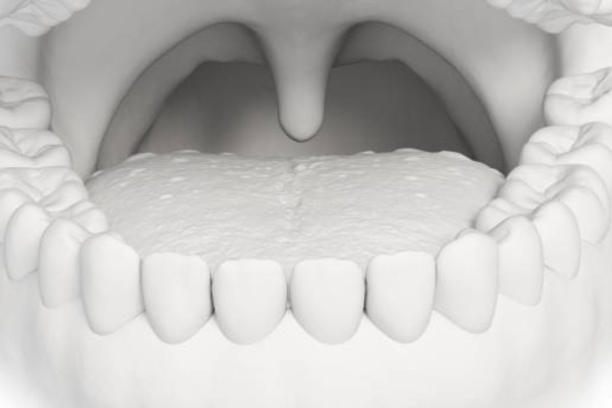
A torus palatinus is a harmless bony growth in the midline of the hard palate. It feels like a hard, raised ridge or mound on the roof of the mouth and can vary in size. Torus palatinus is essentially a birth defect or developmental variant – not cancerous – and most people never need treatment for it. (Cleveland Clinic notes that torus palatinus “is a harmless, noncancerous, bony growth on the roof of your mouth” and only requires removal if it interferes with speaking, chewing or fitting dentures.)
Other benign palatal cysts and growths include nasopalatine duct cysts (fluid-filled sacs behind the front teeth) and minor salivary gland cysts. Nasopalatine duct cysts are benign fluid-filled bumps in the midline palate (behind the two front teeth); they often cause no symptoms unless they enlarge or become infected.
Similarly, oral mucoceles (mucus retention cysts) can form on the palate after minor trauma to a salivary gland. These typically appear as soft, dome-shaped, bluish or translucent swellings that may come and go. Fibromas (irritation fibromas) are firm, smooth, pink nodules of scar tissue that form in response to chronic irritation (from a sharp tooth or dental appliance).
Oral papillomas (cauliflower-like bumps caused by human papilloma virus) can also arise on the palate; these are benign lumps with a rough surface. Small extra or ectopic teeth can sometimes erupt on the palate (hyperdontia), creating a bump until the tooth comes through.
Infectious causes often create painful or ulcerated lumps rather than firm bumps. For example, a herpes simplex (cold sore) infection may produce clustered blisters or a tender ulceration on the palate. Canker sores (aphthous ulcers) are small, yellow-white ulcers with a red rim that can appear anywhere on the oral mucosa (including the palate) and usually heal on their own. Streptococcal throat infection (“strep throat”) can cause tiny red spots or petechiae on the soft palate.
Oral thrush (candida fungus) typically appears as white patches but can give the mouth a “bumpy” or coated feel; it is most common in people with weakened immunity or who use inhaled steroids. Any trauma (such as a burn from hot food or a cut from sharp foods) can cause a tender swelling or blister on the palate that usually heals in days to weeks.
Tumors and neoplasms are less common. Most salivary gland tumors on the palate are benign (e.g. pleomorphic adenomas) and grow slowly, feeling like a soft, movable lump. However, a persistent, new, or rapidly growing lump – especially if hard or ulcerated – raises concern for oral cancer.
Oral cancer (usually squamous cell carcinoma) can occur on the palate. It often presents as a non-healing ulcer or a mass that may feel firm or gritty. Early signs may include a red or white patch (leukoplakia/erythroplakia) or a painless lump that slowly enlarges. Risk factors for cancer include tobacco, heavy alcohol use, and HPV infection, but cancer can occur in anyone.
Distinguishing Benign vs. Concerning Lumps
Most palatal lumps are benign and stable. Benign lesions (like a torus or fibroma) tend to be painless, well-defined, and slow-growing. For instance, a torus is firm and bony and has usually been present for years without change. Benign cysts and mucoceles typically feel soft or fluctuant and may temporarily enlarge or rupture but then heal.
By contrast, warning signs suggest a potentially serious problem. These include a lump or sore that grows or changes over time, especially over weeks; feels hard or fixed; bleeds easily; or shows color changes (such as white or red patches over the lesion). Other red flags are new pain, numbness, difficulty swallowing or speaking, and unexplained weight loss.
In oral cancer, for example, symptoms often include sores or lumps that bleed and do not heal in two weeks. Painless red/white spots (leukoplakia/erythroplakia) that persist may also be precancerous. In short, any palatal lump that is new, growing, ulcerated, or accompanied by bleeding or systemic symptoms warrants prompt evaluation.
Associated Symptoms

Pay close attention to associated symptoms. Pain or tenderness may accompany infected or ulcerated lesions (like canker sores or abscesses) but can also appear in malignancy as the tumor invades tissue. Bleeding from a lump or ulcer that is not due to trauma is concerning. A lump with color change (white or red patches) on or around it can signal dysplasia.
Difficulty chewing, swallowing, or fitting dentures, or a lump that suddenly interferes with these functions, should be noted. Sometimes even ear pain or a persistent sore throat can be related to an oral lesion. Because these symptoms can overlap, self-examination and tracking changes is important.
When to Seek Care
You should see a healthcare professional (dentist, oral surgeon, or primary doctor) if a palatal lump:
-
Persists or worsens for more than 1–2 weeks.
-
Grows in size, changes in shape or color, or bleeds without clear cause.
-
Causes significant pain, numbness, or difficulty swallowing or opening the mouth.
-
Is associated with systemic signs like fever, unexplained weight loss, or a “foul taste” in the mouth.
-
Interferes with dentures or bite alignment.
In practice, many dentists include an oral cancer screening as part of routine checkups. They can often spot suspicious lesions or asymmetries during exams. As one patient resource notes, any new or unusual growth in the mouth should be reported to a dentist or doctor immediately. Early evaluation is especially important for at-risk individuals (tobacco users, heavy drinkers, or those with a history of HPV).
Diagnosis
Diagnosing a palatal lump typically begins with a clinical exam. A dentist or doctor will inspect and palpate the lesion, noting its size, texture, and exact location. They will review your medical history and risk factors. Because many lumps have characteristic appearances (for example, a bony torus is hard and midline, while a mucocele is soft and bluish), often a professional can make a likely diagnosis on exam alone.
Imaging studies may be used to further evaluate the lump. For example, a dental X-ray or CT scan can distinguish a cystic (fluid-filled) lesion from a solid mass. If a nasopalatine duct cyst or other bony growth is suspected, imaging will show a well-defined radiolucent area. Biopsy is the definitive test for any lesion that is unclear or looks suspicious. A small tissue sample (via a needle biopsy or surgical punch) is sent to pathology. According to guidelines, any persistent white or red patch or unexplained ulcer should be biopsied to rule out dysplasia or cancer.
In practice, persistent palatal lumps are often referred to an oral and maxillofacial surgeon or ENT specialist. They may also use techniques like brush cytology or endoscopy (to check deeper structures) as needed. In short, diagnosis relies on combining clinical appearance with targeted imaging and biopsy when indicated.
Treatment
Treatment of a palatal lump depends entirely on its cause:
-
Benign bony growths (Torus palatinus): No treatment is needed if asymptomatic. If a torus interferes with eating, speaking, or denture fit, it can be surgically removed by an oral surgeon. Recovery takes weeks of a soft diet and oral hygiene care.
-
Mucoceles/Fibromas/Other benign lesions: These are often excised under local anesthesia if bothersome. For instance, a persistent mucocele is surgically removed along with adjacent salivary tissue. An irritation fibroma is simply cut out. After removal, pathology will confirm the diagnosis (especially important for any growth that has atypical features). Recurrence is uncommon if the entire lesion (and source of irritation) is removed.
-
Viral/bacterial infections: Herpes (cold sore) outbreaks on the palate may be treated with oral or topical antivirals (e.g. acyclovir). Bacterial infections (such as abscesses) require antibiotics or drainage. Thrush (oral candidiasis) is treated with antifungal lozenges or rinses. Canker sores usually heal on their own in 1–2 weeks; symptomatic relief (topical anesthetics or steroids) may be offered.
-
Benign tumors (e.g. papilloma, pleomorphic adenoma): These are removed surgically, both for symptom relief and to prevent potential growth. Human papilloma virus–related papillomas are typically excised. Salivary gland tumors like pleomorphic adenomas are also excised because, although benign, they can recur or (rarely) transform into malignancy. In all these cases, pathology will verify the tumor type.
-
Precancerous lesions: White or red patches (leukoplakia/erythroplakia) or nonhealing ulcers usually need biopsy and often surgical removal. Complete excision is the safest approach, since these lesions carry a risk of progressing to cancer.
-
Oral cancer: Treatment is multimodal. The mainstay is surgery to remove the tumor, often with a margin of normal tissue. For palate cancers, this may involve partial removal of the hard palate (maxillectomy). Surgery is usually followed by radiation therapy to kill any residual cancer cells. Some patients also need chemotherapy (drug therapy) or newer targeted/immunotherapies, depending on the stage and location. Early-stage cancers confined to the palate may be cured by surgery alone; advanced cancers require combined treatments. For all cancers, follow-up care (nutrition support, speech therapy, regular exams) is crucial.
-
Supportive care: For any palatal lesion, good oral hygiene is important. Removing sources of chronic irritation (such as sharp teeth or ill-fitting dentures) can prevent reactive fibromas. Dentists may smooth or adjust teeth and appliances. Pain from ulcers or surgery can be managed with appropriate analgesics.
In summary, bumps on the roof of the mouth have many causes. Most are benign and easily treated or observed, but any lump that persists, grows, or is accompanied by alarming symptoms (bleeding, ulceration, pain, or weight loss) should be promptly evaluated. A dentist or doctor can often distinguish harmless lumps (like a palatal torus) from lesions that need biopsy, ensuring timely treatment.